Molecular Modeling, Computer-aided drug design & In vitro cell-based assays
Structure-based drug discovery is central to the efficient development of therapeutic agents and to the understanding of metabolic processes. The traditional way of drug discovery is the experimental screening of large libraries of chemicals against a biological target (high-throughput screening or HTS) for identifying new lead compounds. The application of rational, structure-based drug design is proven to be more efficient than the traditional way of drug discovery since it aims to understand the molecular basis of a disease and utilizes the knowledge of the three-dimensional (3D) structure of the biological target in the process. State of the art structure-based drug design methods include virtual screening and de novo drug design; these serve as an efficient, alternative approach to HTS.
By using computational methods and the 3D structural information of the protein target, we are now able to investigate the detailed underlying molecular and atomic interactions involved in ligand:protein interactions and thus interpret experimental results in detail. The use of computers in drug discovery bears the additional advantage of delivering new drug candidates more quickly and cost-efficiently. Computer-aided drug discovery has recently had important successes: new ligands have been predicted along with their receptor-bound structures and in several cases the achieved hit rates (ligands discovered per molecules tested) have been significantly greater than with experimental high-throughput screening.
The central theme of our work at the Biomedical Research Foundation of the Academy of Athens (BRFAA) concerns the computational assessment of the structure and the binding free energy of a ligand-protein complex, the de novo design of novel ligands, the investigation of protein or ligand mutational effects on the free energy of binding, the optimization of potent ligands for a given protein target to enhance activity, the dynamics of the ligand:protein complexes, as well as predicting pharmacokinetic properties of potential leads. Our work is strongly coupled to experimental studies, such as chemical synthesis, in vitro assaying, pharmacokinetic evaluation of potent compounds, and in vivo testing on animal models. Such experimental studies are conducted as a collaboration with research groups at BRFAA or other leading Institutes in Greece and abroad.
___________________________________________________________________________________
ChemBioServer: A platform for filtering, clustering, and visualization of chemical compounds used in drug discovery.
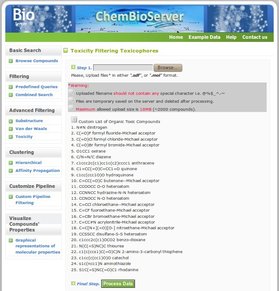
ChemBioServer is a publicly available web application for effectively mining and filtering chemical compounds used in drug discovery. It provides researchers with the ability to (i) browse and visualize compounds along with their properties, (ii) filter chemical compounds for a variety of properties such as steric clashes and toxicity, (iii) apply perfect match substructure search, (iv) cluster compounds according to their physicochemical properties providing representative compounds for each cluster, (v) build custom compound mining pipelines and (vi) quantify through property graphs the top ranking compounds in drug discovery procedures. ChemBioServer allows for pre-processing of compounds prior to an in silico screen, as well as for post-processing of top-ranked molecules resulting from a docking exercise with the aim to increase the efficiency and the quality of compound selection that will pass to the experimental test phase.
You can use ChemBioServer here: http://bioserver-3.bioacademy.gr/Bioserver/ChemBioServer/
A full description of the platform can be found here:
ChemBioServer: a web-based pipeline for filtering, clustering and visualization of chemical compounds used in drug discovery.
Athanasiadis E, Cournia Z, Spyrou G. Bioinformatics. 2012 Nov 15;28(22):3002-3. Epub 2012 Sep 8.
http://dx.doi.org/10.1093/bioinformatics/bts551
___________________________________________________________________________________________
Insights
into the mechanism of oncogenesis of the mutated PI3Kα and application to
rational drug design
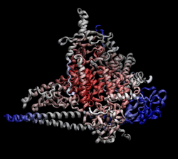
The PI3Kα kinase is involved in fundamental cellular processes such as cell proliferation and differentiation. Somatic mutations within the gene PIK3CA, which encodes the catalytic subunit of PI3Kα (p110α), are frequently observed in a variety of human tumors, including breast, colon, endometrial cancers and glioblastomas. These mutations are scattered over the length of p110α, but two hotspots account for nearly 80% of them: an H1047R substitution close to the C-terminus, and a cluster of three change-reversal mutations (E542K, E545K, Q546K) in the helical domain of p110α. Both types of these mutations increase the kinase activity of the enzyme, upregulate the downstream AKT pathway and VEGF signaling, and thus stimulate cell transformation, tumorigenesis and angiogenesis. Mutated proteins such as PI3Kα can drive tumor progression due to conformational changes that result in upregulated enzymatic activity. Understanding how these mutations cause tumorigenesis is central to developing new therapeutics for cancer.
Our first goal in this project is to gain insights into the oncogenic mechanism of two commonly-expressed PI3Kα mutants (H1047R and E545K). Understanding how these “hotspot” mutations lead to the increased PI3Kα activity is paramount to developing new treatments for cancer. To this end, we perform extensive Molecular Dynamics (MD) simulations of the wild-type and mutant forms of PI3Kα and monitor the differences between them by means of structural, thermodynamic and kinetic properties. A second aim of the project is to design novel inhibitors that will selectively inhibit the two mutants (E545K and H1047R) and not their wild-type counterpart, which could result in the absence of adverse effects. This computational study is complemented by several experimental groups from BRFAA and other leading research Institutes in the country regarding the synthesis of potential leads, in vitro/in vivo assaying and in vivo testing using animal models. Following the assays, potent compounds are iteratively optimized for both pharmacological and structural properties with computational methods and re-assayed for enhanced activity.
Mutant H1047R PI3Kα crystal structure (PDB ID: 3HIZ)
colored by B factor (Bfactor-max=126 Å2 (blue),
Bfactor-min=30 Å2 (red).
___________________________________________________________________________________
Interaction of aminoadamantine derivatives with M2TM
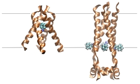
The M2 protein of influenza A virus is a pH-activated membrane-spanning proton channel that plays an important role in the acidification and uncoating of the endosome-entrapped virus and in viral assembly. At low pH, it adopts an open conformation, whereas at high pH it forms a closed pore. The membrane-bound M2 peptide bundle (M2TM) consists of four helices and is the target of amantadine and rimantadine, two anti-influenza drugs. Resistance to these drugs has reached more than 90%. It is thus important to design novel therapeutic molecules to target the M2TM channel. We are interested in designing small molecules with inhibitory action against the M2TM channel, using docking simulations and Free Energy Perturbation (FEP) calculation. Moreover, promising candidates will be further studied by means of Molecular Dynamics simulations. This project is in collaboration with Prof. Antonios Kolocouris lab, Department of Pharmaceutical Chemistry, Athens University.
Snapshop of M2TM with bound amantadine in low
pH (left) and high pH (right) [Image: Evi Gkeka]
Snapshop of M2TM with bound amantadine in low
pH (left) and high pH (right) [Image: Evi Gkeka]
___________________________________________________________________________________
Mechanistic and inhibition studies of the Arp2/3 complex activation
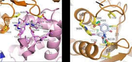
Precisely controlled polymerization of actin is required for a broad range of cellular processes from lamellar motility to organelle transport. The Arp2/3 complex has emerged as a key nucleator of branched actin filaments because of its ability to bind existing filaments and initiate new filament growth. New filament growth is preceded by an activation of the Arp2/3 complex that is believed to be the result of a large conformational change. Despite the essential roles of the Arp2/3 complex in vivo, the exact mechanism of its activation is not known. Our goal is to unravel the detailed mechanism of Arp2/3-mediated branching nucleation in order to aid studies of the in vivoregulation of the Arp2/3 complex. We study the Arp2/3 mechanism with Steered Molecular Dynamics and collaborate with experimental groups (Nolen lab, University of Oregon, USA) to confirm our findings.
Apart from the involvement of the Arp2/3 complex into actin filament nucleation, the complex also serves other important in vivo functions such as the formation of podosomes in macrophages and in endocytic actin patch motility in yeast. It is also implicated in actin remodeling processes, such as grown cone pathfinding in neurons and lamellar motility in keratocytes. However, the contribution of the Arp2/3 complex in all of the above-described functions has been the subject of dispute for years, mainly because of the lack of simple methods to inhibit the Arp2/3 complex reversibly in vivo. In order to be able to efficiently inhibit the Arp2/3 complex in living cells so that important in vivo functions of the complex can be monitored, we aim to advance small molecule inhibitors of the Arp2/3 complex. To achieve this, we optimize recently-discovered small molecules in order to improve their inhibition potency in living cells. We also employ virtual screening techniques to discover novel inhibitors. Selected inhibitors will be tested first in in vitro assays and their efficacy will then be monitored in vivo by the collaborating Nolen lab. When the desired potency is reached, the crystal structure of the inhibitor within the Arp2/3 complex will be pursued by the Nolen lab.
Apart from the involvement of the Arp2/3 complex into actin filament nucleation, the complex also serves other important in vivo functions such as the formation of podosomes in macrophages and in endocytic actin patch motility in yeast. It is also implicated in actin remodeling processes, such as grown cone pathfinding in neurons and lamellar motility in keratocytes. However, the contribution of the Arp2/3 complex in all of the above-described functions has been the subject of dispute for years, mainly because of the lack of simple methods to inhibit the Arp2/3 complex reversibly in vivo. In order to be able to efficiently inhibit the Arp2/3 complex in living cells so that important in vivo functions of the complex can be monitored, we aim to advance small molecule inhibitors of the Arp2/3 complex. To achieve this, we optimize recently-discovered small molecules in order to improve their inhibition potency in living cells. We also employ virtual screening techniques to discover novel inhibitors. Selected inhibitors will be tested first in in vitro assays and their efficacy will then be monitored in vivo by the collaborating Nolen lab. When the desired potency is reached, the crystal structure of the inhibitor within the Arp2/3 complex will be pursued by the Nolen lab.
Modeling of membrane-associated systems
Biological membranes are crucial to cell life as they perform a variety of functions. The plasma membrane, which is the outer cell membrane, encloses the cell, defines its boundaries, senses external signals, serves as a selective barrier for water and ions, and plays an important role in signal transduction. Despite their many different functions, all biological membranes have a common basic structure: the lipid bilayer. Cell membranes are dynamic, fluid structures where the molecules are able to diffuse rapidly in the plane of the membrane. Membranes of eukaryotic cells have a complex composition consisting of hundreds of different lipids and cholesterol, plus different proteins attached to or associated to the membrane.
More than half of all proteins interact with membranes. Membrane-associated proteins can be integral (i.e., embedded in the membrane), such as the influenza virus M2 proton channel (M2TM protein), or peripheral (i.e., interact with the membrane non-covalently), such as the PI3Kα, which is a lipid kinase recruited to the membrane to phosphorylate the substrate PIP2 to PIP3. Both of these proteins represent important pharmaceutical targets. M2TM is integral in the viral envelope and its inhibition leads to blocking viral replication. PI3Kα is implicated in extraordinarily diverse cell functions such as proliferation, cell growth and differentiation, and has been recently found to be mutated in many cancers. Understanding how the membrane influences the structure and function of these proteins is key to designing novel inhibitors.
Biological membranes come to contact not only with naturally-occurring entities, such as proteins, but also with man-made entities, such as drugs or nanoparticles, that need to surpass this barrier in order to enter the human cells. The increasing applications of nanotechnology in medicine increase the likelihood that engineered nanomaterials, such as diagnostic and therapeutic nanoparticles (NPs), will come in contact with human cells. Therefore, the first step into assessing the NP cytotoxicity requires a thorough understanding of the NP-membrane interaction mechanism. We work on associating nanoparticle characteristics with specific membrane interaction patterns with and design NPs with tailored functionalities, such as direct cellular entry.
___________________________________________________________________________________
Designing nanoparticles with tailored functionalities
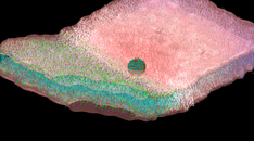
The use of nanoparticles (NPs) of variable chemistry and architecture is one of the new promising directions in gene delivery and silencing and cellular imaging. Moreover, the development of therapeutic nanoparticles (TNPs) for cancer imaging and treatment has shown excellent promise for drug delivery. However, despite the extensive research on NP applications in therapeutics, there are only a limited number of NP systems that have been clinically tested. Before NPs contact healthy human cells, their cytotoxicity needs to be further assessed and quantified. The entry point of a NP to a cell is the plasma membrane. Therefore, the first step into assessing the NP cytotoxicity requires a thorough understanding of the NP-membrane interaction mechanism.
The main aim of this project is to systematize how NP characteristics, such as size, charge and surface attributes, including peptide coating, affect the interaction of the NP with the cell membrane as well as how they induce different internalization mechanisms. We perform extensive Coarse-Grained Molecular Dynamics (CGMD) simulations and free energy calculations to gain insights into the mechanism of NP-membrane interaction with at the microscopic level. Multiple biophysicochemical attributes of the system, such as stretching, elasticity and thermal fluctuations of the membrane, as well as the free energy release at the contact site, are monitored. Our ultimate goal is to associate NP characteristics with specific membrane interaction types and design NPs with tailored functionalities, such as direct cellular entry. Moreover, we aim to provide insights of the NP aggregation and peptide coating mechanisms.
This project is in collaboration with Dr. Panagiotis Angelikopoulos, ETH, Zurich.
Protein Structure Prediction with Homology and Loop Modeling
The unknown 3D structure of a query protein (or parts of it) can be predicted by homology modeling, namely from their amino acid sequence and an experimental structure of a homologous protein (template). Models based on templates with more than 50% sequence identity are generally very accurate and can exhibit ~1 Å atom RMSD from the native structure. Proteins with 30-50% sequence identity share at least 80% of their structures, those with 20-30% sequence identity share at least 55%, and when sequence identity is lower than 20% proteins may display 30% or even lower structural similarity. However, there are cases in nature where proteins with no evident sequence similarity share the same fold. Protein threading implements knowledge-based scoring functions to identify potential templates that fall into this category (fold recognition), and use them to build models of the query protein. Protein threading performs well when sequence identity drops below 30%, the region where Homology Modeling cannot generate good predictions. Finally, when fold recognition fails, then the structure can be predicted ab initio, namely from the sequence alone using physical principles rather than (directly) previously solved structures. Ab initio structure prediction is more computationally demanding than homology modeling and protein threading, and has a prediction limit of ~200 amino acids. Loop modeling falls into this category. The best performing programs in this category produce models with resolution ~ 5 Å, hence ab initio structure prediction should be used as the last resort.
Force Field Development
The functional form of the force field used in a MD simulation must be used in conjunction with a set of empirical parameters, which are molecule dependent and must be optimized prior to performing simulations. This optimization step is generally referred to as parameterization of the force field. The reliability of a molecular mechanics (MM) calculation is dependent on both the functional form of the force field and the numerical values of the associated parameters. Thus, the first necessary step towards a reliable MD simulation is the parameterization procedure. Most all-atom empirical force fields used in common MD packages (such as CHARMM) are equipped with parameter sets for modelling basic building blocks of biomolecules, which can then be combined for more complex molecular systems. However, for more exotic molecules such as several drug classes a force field is not available. AFMM (Automated Frequency Matching Method) provides an efficient, automated way to generate intra-molecular force field parameters using normal modes. The method can be, in principle, used with any atom-based molecular mechanics program, which has the facility of calculating normal modes and the corresponding eigenvectors. The basic principle behind the AFMM method is to iteratively tune an initial MM parameter set in order to reproduce the normal modes generated from a quantum mechanical (QM) calculation. The program refines an initial parameter set, which can either be a pre-existing set or using chemically-reasonable estimation.
You can request the program code and manual by sending an email to Zoe Cournia.
Apart from the intra-molecular force field parameters (i.e., force constants), partial atomic charges, which is not an experimentally-observable quantity, need to be calculated. The electrostatic properties of a molecule are a consequence of the electron and the nuclei distribution. Thus, it is reasonable to assume that one should be able to obtain a set of partial atomic charges using quantum mechanics. However, as mentioned the partial atomic charge is not an experimentally observable quantity and cannot be unambiguously calculated from the wavefunction. Many methods have been proposed for calculating the partial atomic charges from quantum mechanics. The most commonly used in our studies is the CHELPG method, in which the set of partial atomic charges that best reproduces the quantum mechanical electrostatic potential at a series of points surrounding the molecule is derived.
